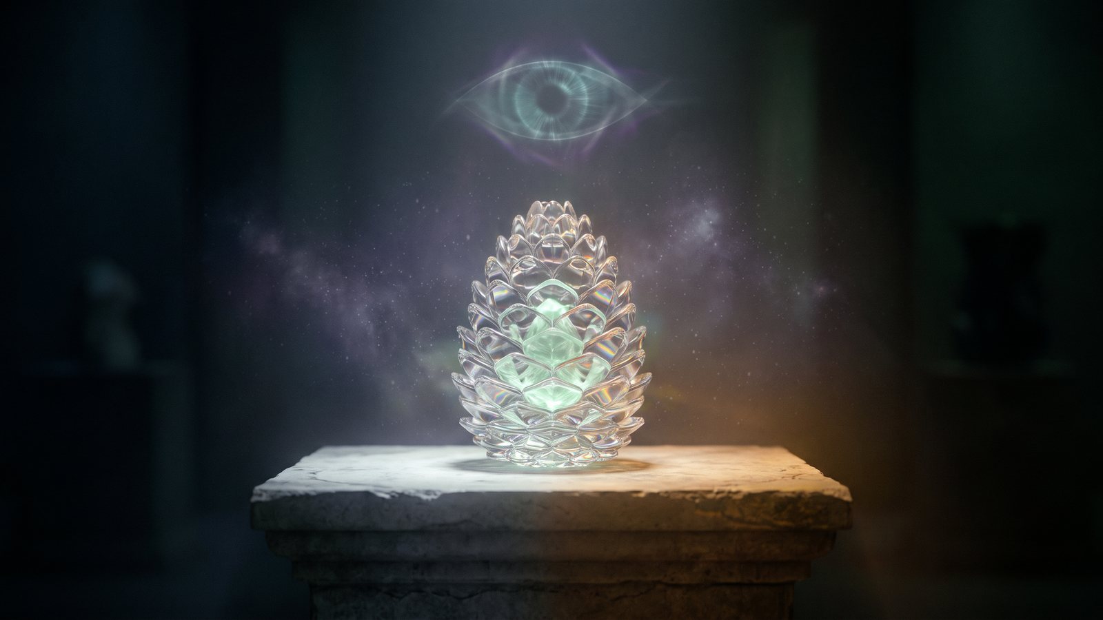
THE SHROOMVERSE JOURNAL ✳ 16 MIN READ
The pineal gland and the “third eye”: what the gland really does, and which claims hold up
Topic: DMT
Published
Listen: the 3-minute version
AI-narrated summary of this article
Short answer, checked 21 October 2026
The pineal gland “third eye” is a metaphor, not human anatomy: the human pineal gland cannot sense light and does not see. Its established job is making melatonin at night, which helps time the body’s daily rhythm. The “third eye” label has three roots: Descartes calling the gland the principal seat of the soul in the 1640s; 19th-century anatomists who saw it as a vestige of a real light-sensing eye on top of the head, which the tuatara and many lizards still have; and Theosophy, which in 1888 recast it as an organ of spiritual vision. Pineal calcification is common, and fluoride does build up in the gland, but no study shows that any “activation” or “decalcification” practice opens anything.
THE SHORT VERSION · SWIPE


On this page 10 sections
Search for the pineal gland and you will find two very different organs. One is a small hormone gland described in endocrinology textbooks. The other is a “third eye” that can be calcified, detoxed, activated or opened. This article sets out what the first one does, how the second one came to exist, and what the studies people cite actually measured.
The DMT side of the story already has its own fact-checks. In short: DMT has never been measured being made by a human pineal gland, and in rats with the gland removed, DMT was still present in the cortex at levels no lower than in rats with it (is DMT in the pineal gland?). And no study has measured DMT during meditation or breathwork (can breathwork or meditation release DMT?). For DMT itself, start with what is DMT.
Education only, not guidance
This article describes published research and history. It gives no instructions and recommends no practice, diet, supplement or product for the pineal gland. If you have concerns about sleep or a finding on a brain scan, speak to a doctor. Evidence labels used below: established (shown in peer-reviewed studies and not seriously disputed), preliminary (early, small, animal-only, cross-sectional or indirect) and unresolved (an open question).
What is the pineal gland, and what does it do?
The pineal gland is a small, cone-shaped gland near the centre of the brain, between the two hemispheres (Wada et al., 2026). The adult gland weighs less than a fifth of a gram, and its principal function is producing melatonin, a hormone that regulates circadian rhythm, the body’s roughly 24-hour cycle (Nichols, 2018). Evidence: established.
Melatonin is a night signal. Pineal melatonin is always produced during the night, and how long the nightly release lasts depends directly on the length of the night, so it tells the body both the time of day and the season (Cipolla-Neto and Amaral, 2018). Because it is normally secreted only in the dark, its rhythm is widely used as a marker of the internal clock. Melatonin is also used as a treatment for some circadian sleep disorders, such as jet lag and delayed sleep phase (Arendt and Aulinas, Endotext). Evidence: established. That last point is a description of the literature, not a recommendation.
If it cannot see, how does the pineal gland know it is dark?
Through the eyes. In mammals the pineal gland “does not detect light by itself”: it serves a neuroendocrine (hormone-signalling) role, secreting melatonin at night (Romero and de Souza, 2025). The light-sensing ability that pineal organs have in many other vertebrates is thought to have been lost in mammals, along with much of the organ’s size, a shift the authors of a 2026 review describe as a move from direct photoreception to hormonal regulation of circadian rhythms (Wada et al., 2026). The gland receives its information about the light-dark cycle from the environment, by way of the nervous system (Arendt and Aulinas, Endotext). Evidence: established.
Why is the pineal gland called the third eye?
Three separate ideas fed the name, centuries apart. Only one of them is biology, and it applies to lizards, not people.
- AntiquityPharaonic Egypt is said to have equated the gland with the eye of Horus; later traditions treat it as the seat of the soul or third eye.Source Nichols, 2018
- 1640–1649Descartes calls the gland the principal seat of the soul, in letters and in The Passions of the Soul (1649).Source Stanford Encyclopedia of Philosophy
- Late 1800sSeveral scientists independently propose that the pineal is a vestige of a dorsal (top-of-the-head) third eye.Source Stanford Encyclopedia of Philosophy
- 1888Helena Blavatsky, founder of Theosophy, recasts that vestige as an atrophied “organ of spiritual vision”.Source Stanford Encyclopedia of Philosophy; Paditz et al., 2025
- 1958Melatonin, the gland’s hormone, is first isolated.Source Stanford Encyclopedia of Philosophy
- 2000 onwardThe gland is popularly linked to DMT, after a speculation in Rick Strassman’s book.Source Our Strassman and pineal articles
Descartes: the “principal seat of the soul”
René Descartes regarded the pineal gland as the principal seat of the soul and the place in which all our thoughts are formed. His main reason was anatomical: he could not find any other part of the brain that is not double, and since we have one thought at a time from two eyes and two ears, he reasoned the impressions must unite in a single structure. Only a few people accepted his pineal theory while he was alive, and it was almost universally rejected after his death (Stanford Encyclopedia of Philosophy). Evidence: established as history.
Some of his anatomy was wrong even by the standards of his time. He thought the gland hung in the middle of the brain’s fluid-filled ventricles and was fed by small arteries, but Galen had already pointed out that it is not suspended there and is surrounded by veins rather than arteries (Stanford Encyclopedia of Philosophy). A 2025 history paper calls the seat-of-the-soul view “a famous error” that critics from Thomas Willis to Kant rejected, and that still circulates today (Paditz et al., 2025). Evidence: established as history.
Theosophy: from anatomy to “spiritual vision”
The modern spiritual meaning comes mostly from the late 19th century. As comparative anatomists began describing the pineal as a vestige of a dorsal third eye, Helena Blavatsky, the founder of Theosophy, identified that “third eye” with the “eye of Shiva” of Hindu mystics and concluded, in her 1888 book The Secret Doctrine, that the human pineal body is an atrophied vestige of an “organ of spiritual vision” (Stanford Encyclopedia of Philosophy). The 2025 history paper adds that she combined it with ideas about chakras, and that Rudolf Steiner later linked the gland’s calcium deposits to cosmic energies (Paditz et al., 2025). Evidence: established that these ideas were published; the ideas themselves are beliefs, not findings.
Is there a real third eye in any animal?
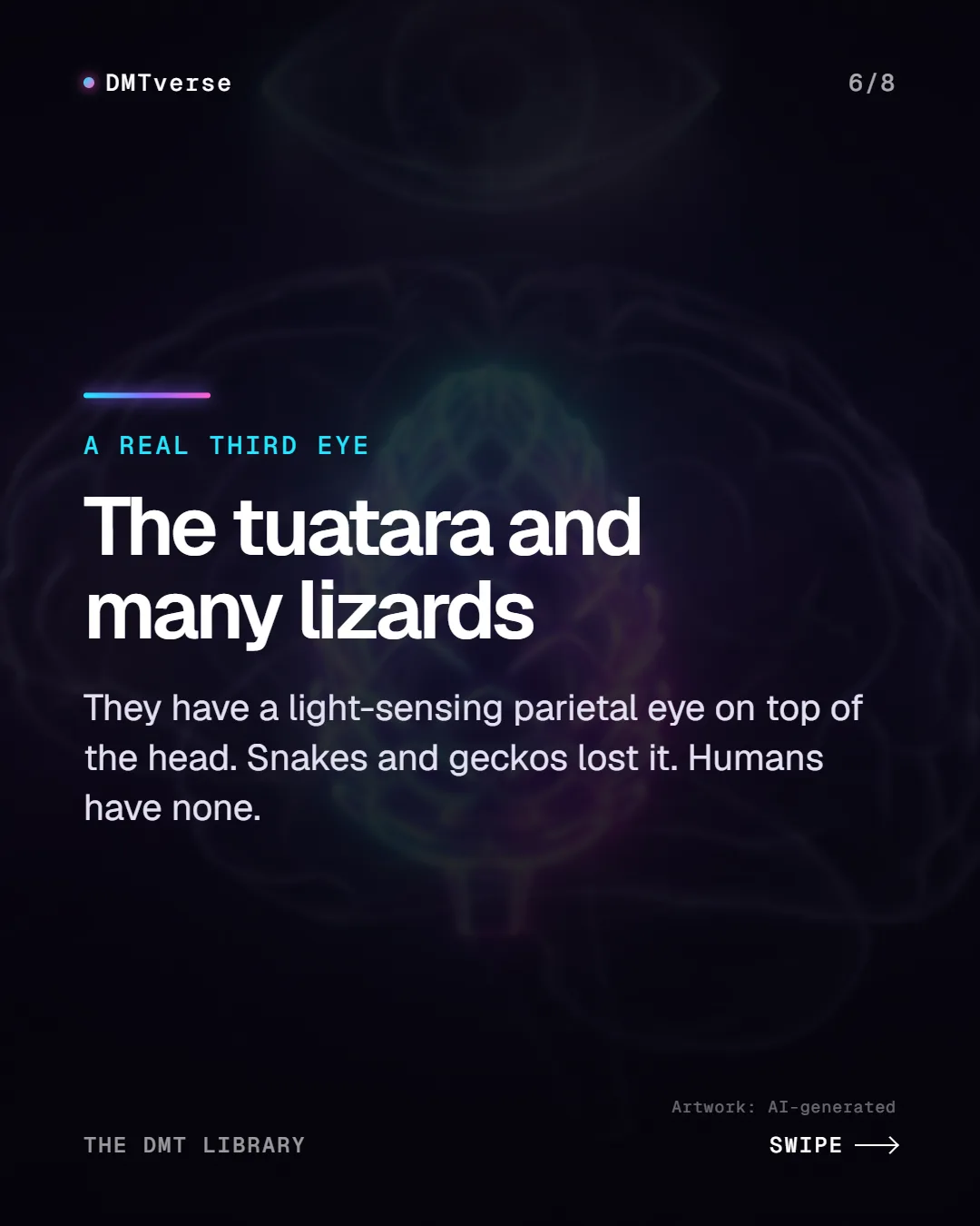
Yes, and it explains why the anatomists’ idea was reasonable. Many lizards and the tuatara, a lizard-like reptile, have a parietal eye: a structure derived from the pineal complex that contains a simplified retina with light-sensing cells. It sits in an opening in the top of the skull, the parietal foramen, and the tuatara’s has a retina and a lens. It cannot form images (Romero and de Souza, 2025). Evidence: established.
It is not universal, even in reptiles. Snakes and geckos have lost the parietal eye, as have turtles, crocodilians and birds (Romero and de Souza, 2025). In lizards that have one, its light-sensing cells contain two opposing signalling pathways in the same cell, one most sensitive to blue light and one to green (Su et al., 2006). Experiments that cover or remove it alter how long lizards bask in the sun, their body temperature and their daily activity patterns (Romero and de Souza, 2025). Evidence: established. Humans have no parietal eye.
What is pineal gland calcification?
A calcified pineal gland is one containing mineral deposits, sometimes called “brain sand” (acervuli). The deposits are mostly hydroxyapatite, the main mineral in bone, and a 2018 review notes the gland has the highest calcification rate of any organ or tissue in the human body (Tan et al., 2018). The deposits are so common that they make the gland an easily identified reference point on brain X-rays (Stanford Encyclopedia of Philosophy). Evidence: established.
How common it is depends heavily on age and on the imaging method:
- Children: on head CT scans of 242 patients under 16, calcification appeared in 1% of those younger than 6, 8% of those younger than 10, and 39% of those aged 8 to 14 (Doyle and Anderson, 2006).
- Hospital CT, adults and teenagers: in 1,614 patients over 15 at a Thai teaching hospital, 67.0% had pineal calcification (Kitkhuandee et al., 2014).
- Dental cone-beam CT: in scans from 500 patients referred for implant treatment, prevalence was 58.8%, with no difference between women and men (Mutalik and Tadinada, 2017).
- Plain skull X-rays: in 480 patients aged 3 to 89 in Iraqi Kurdistan, the rate was 26.9%, rising after the first decade of life (Mohammed et al., 2016).
These samples are hospital or clinic patients, not the general population, and CT picks up far smaller deposits than plain X-ray, which is the likeliest reason the figures differ so much. Evidence: established that calcification is common and increases with age; exact population rates are preliminary.
Does a calcified pineal gland make less melatonin?
Possibly. In a 1999 study of 26 people, the estimated volume of uncalcified pineal tissue on CT was positively associated with a melatonin breakdown product collected in urine over 24 hours, and the authors argued that the age-related fall in melatonin could be explained by increasing calcification (Kunz et al., 1999). Calcification has also been statistically associated with stroke in the Thai CT study (Kitkhuandee et al., 2014). These are associations, not proof of cause. Evidence: preliminary.
Does fluoride calcify the pineal gland?
Fluoride does accumulate there; whether that harms the gland in people is not established. The study most often cited is Jennifer Luke’s, published in 2001. She measured fluoride in the pineal gland, muscle and bone of 11 aged cadavers. Pineal fluoride correlated with pineal calcium (r = 0.73), but not with bone fluoride, and by old age the gland’s fluoride-to-calcium ratio was higher than bone’s (Luke, 2001). Evidence: established as a finding in 11 elderly cadavers; it measured accumulation, not function.
What that study did not show matters as much. It did not measure melatonin, sleep or any symptom, and 11 bodies cannot show what causes calcification. Luke’s related animal work was a 1997 doctoral thesis (Cunningham et al., 2021, reference list), which we have not read. A 2018 review lists fluoride as one possible environmental contributor to early calcification, citing Luke’s study and a bird study (Tan et al., 2018). Evidence: unresolved for any effect on the human gland’s function.
The only Canadian data we found are indirect. Using the 2012–2013 Canadian Health Measures Survey, researchers linked higher tap-water fluoride to a 34% higher relative risk of reporting less sleep than recommended in one analysis, though the survey-weighted estimate was less precise. Urinary fluoride was not linked to sleep duration, and neither measure was linked to sleep problems or daytime sleepiness (Cunningham et al., 2021). The design was cross-sectional and sleep was self-reported. The authors call their results hypothesis-generating and say the direction of the association is not established. Evidence: preliminary.
Does “pineal gland activation” or opening the third eye work?
There is no evidence that it does, and part of the claim rests on a misunderstanding. A healthy pineal gland is already active: it switches on melatonin production every night without any technique (Cipolla-Neto and Amaral, 2018). And the human gland has no light-sensing cells to “open”; its information about light arrives indirectly (Romero and de Souza, 2025). Evidence: established for the physiology.
On “how to open the third eye” and “pineal decalcification” methods (diets, supplements, sound frequencies, crystals, sungazing and so on), we searched Europe PMC on 21 October 2026 and found no controlled human study showing that any of them reduces pineal calcification, raises melatonin through the pineal, or produces any perception that could be called a third eye. Absence of trials is not proof that nothing happens, but it means the claims are untested, not proven. Evidence: unresolved. We describe none of these practices here.
One scientific paper goes further than the evidence on its own terms. The 2018 calcification review closes by suggesting that pineal health might be preserved by slowing premature calcification or even “rejuvenating” the calcified gland (Tan et al., 2018). That is a hypothesis in a review; it reports no human trial of such a treatment. Meditation and breathwork are covered in our breathwork and meditation fact-check. Evidence: unresolved.
About “pineal detox” claims
Claims that a diet, supplement, sound or course can decalcify or activate the pineal gland are claims that, as far as we could find, no controlled human study has tested. Pineal calcification is usually an incidental finding on a scan. If a scan report mentions it, or you have ongoing sleep problems, ask a doctor what it means for you.
How did the pineal gland get linked to DMT?
The DMT link is the newest layer. Psychiatrist Rick Strassman’s group studied melatonin before DMT, and his 2000 book proposed, as an openly speculative idea, that the pineal gland releases DMT at moments such as birth and death. Our Rick Strassman study article covers that history. A 2018 review lists DMT secreted by the pineal at birth, during dreaming and near death among the ideas the scientific evidence does not support (Nichols, 2018). Evidence: established that the idea was proposed as a speculation.
Two measurements anchor the fact-check. In Strassman’s own 1994 controlled study of 11 volunteers, DMT raised several hormones but left blood melatonin unaffected (Strassman and Qualls, 1994). In 2019, rats whose pineal gland had been removed still had DMT in the cortex at levels similar to rats with the gland (Dean et al., 2019). The full rat evidence is in is DMT in the pineal gland?, the wider question in does the brain make DMT?, and the enzyme story in INMT and DMT. Evidence: preliminary (one small human study; rats).
Pineal gland and third eye: myth vs evidence
| Claim | Verdict | What the evidence shows |
|---|---|---|
| The pineal gland is a human third eye | Myth (metaphor) | The human gland does not detect light by itself (Romero and de Souza, 2025). |
| Some animals have a real third eye | True | The tuatara and many lizards have a light-sensing parietal eye; snakes and geckos lost it (Romero and de Souza, 2025). |
| Descartes called it the seat of the soul | True | He called it the principal seat of the soul; the view was soon rejected (Stanford Encyclopedia of Philosophy). |
| The pineal gland’s job is making melatonin | True | Its principal known function, timing the daily rhythm (Nichols, 2018; Cipolla-Neto and Amaral, 2018). |
| Most adults have a calcified pineal gland | Often true on CT | 67.0% in one hospital CT series; lower on plain X-ray (Kitkhuandee et al., 2014; Mohammed et al., 2016). |
| Fluoride accumulates in the pineal gland | True (small study) | Found in 11 aged cadavers, correlated with calcium (Luke, 2001). |
| Fluoride has calcified your third eye | Unsupported | No study has shown fluoride causes pineal calcification or loss of function in people. |
| You can decalcify or activate it with a technique | Untested | No controlled human trial found; the gland is already active nightly. |
| The pineal gland releases DMT | Unsupported | Never measured in humans; rat brain DMT persisted without it (Dean et al., 2019). |
For more DMT claims tested the same way, see DMT myths, fact-checked and why DMT is called the spirit molecule.
What we still don’t know
- How common pineal calcification is in the general population, rather than in hospital patients, measured the same way everywhere (unresolved).
- Whether calcification itself lowers melatonin or simply tracks ageing; the key study had 26 people (preliminary).
- Whether fluoride at everyday exposure levels affects the human pineal gland’s function at all (unresolved).
- Whether any intervention can reduce pineal calcification in people; we found no trial (unresolved).
- Whether the human pineal gland makes any DMT; it has never been measured (unresolved).
The fair summary: the pineal gland is a real, well-studied hormone gland, and the third eye is a real organ in lizards and the tuatara. Joining those facts to Descartes’ soul and Theosophy’s spiritual vision produced a powerful story, but the human gland does not see, and the activation, decalcification and DMT claims built on that story remain untested or unsupported.
Common questions
Is the pineal gland really a third eye?
Not in humans. The human pineal gland does not detect light by itself; it makes melatonin at night using light information that reaches it indirectly. A real light-sensing “third eye”, the parietal eye, exists in the tuatara and many lizards.
What does the pineal gland do?
Its principal known function is producing melatonin, a hormone released at night that helps time the body’s daily (circadian) rhythm and signals the length of the night.
What hormone does the pineal gland release?
Melatonin. It is normally secreted during darkness, and its rhythm is used by researchers as a marker of the body’s internal clock.
Why is the pineal gland called the third eye?
Because of three ideas: Descartes calling it the principal seat of the soul in the 1640s; late-19th-century anatomists who saw it as a vestige of a dorsal third eye; and Helena Blavatsky, who in 1888 called it an atrophied organ of spiritual vision.
What is a calcified pineal gland?
One containing mineral deposits, mostly hydroxyapatite. It is common and increases with age: 1% of children under 6 in one CT study, and 67.0% of patients over 15 in a large hospital CT series.
Does fluoride calcify the pineal gland?
Not shown. A 2001 study of 11 aged cadavers found fluoride accumulates in the gland and correlates with its calcium. It did not test cause or function. A Canadian survey found a link between tap-water fluoride and short sleep in one analysis, but its authors call it hypothesis-generating.
How do you open your third eye or activate the pineal gland?
There is no evidence that any technique does either. The gland is already active every night, and we found no controlled human study showing any practice decalcifies it or creates third-eye perception. We give no methods.
Does the pineal gland make DMT?
It has never been measured doing so in humans. In rats with the gland removed, DMT was still found in the cortex at levels no lower than in rats with it.
Shroomverse sells mushroom products. This article is general information about the pineal gland and the “third eye” idea. It is not medical, legal or purchasing advice, and it does not recommend any product.
SOURCES
- N,N-dimethyltryptamine and the pineal gland: Separating fact from myth (Nichols DE, Journal of Psychopharmacology 32(1):30–36, 2018), SAGE (peer-reviewed); abstract via Europe PMC. Accessed . Abstract read; DOI checked on Crossref. Supports the eye of Horus, seat-of-the-soul and third-eye traditions; pineal DMT claims at birth, dreaming and near death as not consistent with the evidence; the adult gland weighing less than a fifth of a gram; melatonin production regulating circadian rhythm as its principal function.
- Physiology of the Pineal Gland and Melatonin (Arendt J, Aulinas A, in Endotext, MDText.com; PMID 31841296), Endotext (peer-reviewed online endocrinology textbook), via Europe PMC. Accessed . Abstract read via Europe PMC; the NCBI Bookshelf full text showed a browser check on this date and was not read. Supports the gland receiving light-dark information from the environment and converting it into melatonin secretion; melatonin normally secreted only in darkness and used as a circadian phase marker; melatonin used for some circadian sleep disorders (delayed sleep phase, non-24-hour, jet lag).
- Melatonin as a Hormone: New Physiological and Clinical Insights (Cipolla-Neto J, Amaral FG, Endocrine Reviews 39(6):990–1028, 2018), Endocrine Society (peer-reviewed); abstract via Europe PMC. Accessed . Abstract read; DOI checked on Crossref. Supports pineal melatonin always being produced at night, with the duration of secretion depending on the length of the night, coordinating daily and seasonal physiology.
- Descartes and the Pineal Gland (Lokhorst G-J, Stanford Encyclopedia of Philosophy, Summer 2026 archive edition; first published 2005, revised 2013), Stanford Encyclopedia of Philosophy, Stanford University. Accessed . Archived entry read in full (the current URL now points to this archive). Supports Descartes regarding the gland as the principal seat of the soul; his “not double” reasoning; The Passions of the Soul (1649); errors about the ventricles and arteries already noted by Galen; near-universal rejection after his death; late-19th-century vestigial dorsal third-eye hypothesis; melatonin first isolated in 1958; Blavatsky (1888) and the “organ of spiritual vision”; calcifications as an X-ray landmark.
- The pineal gland as the seat of the soul (René Descartes): History of reception, enlightenment, and consequences of a famous error (Paditz E, Shevchenko O, Upreti K, Journal of the History of the Neurosciences 35(2):109–155, published online 2025), Taylor & Francis (peer-reviewed); abstract via Europe PMC. Accessed . Abstract read; DOI checked on Crossref (online 24 November 2025). Supports early criticism by Willis, Spinoza, Leibniz and Kant; the view still being propagated; Blavatsky adding ideas about chakras in 1888; Steiner linking pineal calcite deposits to cosmic energies in 1922/1923.
- Evolution of Pineal Nonvisual Opsins in Lizards and the Tuatara and Identification of Lepidopsin: A New Opsin Gene (Romero RD, de Souza FSJ, Genome Biology and Evolution 17(5):evaf058, 2025), Oxford University Press (peer-reviewed, open access); full text via Europe PMC, PMC12043008. Accessed . Full text read. Supports the mammalian pineal gland not detecting light by itself and secreting melatonin at night; the parietal eye of many lizards and the tuatara as a pineal-complex structure with a simplified retina, set in the parietal foramen, unable to form images; the tuatara’s parietal eye having a retina and lens; loss in snakes, geckos, turtles, crocodilians and birds; covering or removal altering basking, body temperature and activity patterns.
- Parietal-eye phototransduction components and their potential evolutionary implications (Su CY, Luo DG, Terakita A, et al., Science 311(5767):1617–1621, 2006), American Association for the Advancement of Science (peer-reviewed); abstract via Europe PMC. Accessed . Abstract read; DOI checked on Crossref. Supports two antagonistic light-signalling pathways in the same parietal-eye photoreceptor, one most sensitive to blue and one to green light.
- From pineal photoreception to optogenetics: functional colour opponency based on a pineal bistable opsin (Wada S, Kawano-Yamashita E, Koyanagi M, Terakita A, Eye and Vision 13, 2026), BioMed Central (peer-reviewed, open access); full text via Europe PMC, PMC13094114. Accessed . Full text read (relevant passages). Supports the human pineal as a small cone-shaped structure near the centre of the brain between the hemispheres; pineal-related organs in many vertebrates being called the third eye; pineal photosensitivity thought lost in mammals, reflecting a shift from direct photoreception to neuroendocrine circadian regulation.
- Pineal Calcification, Melatonin Production, Aging, Associated Health Consequences and Rejuvenation of the Pineal Gland (Tan DX, Xu B, Zhou X, Reiter RJ, Molecules 23(2):301, 2018), MDPI (peer-reviewed review, open access); full text via Europe PMC, PMC6017004. Accessed . Abstract and relevant full-text passages read. Supports the gland having the highest calcification rate of any organ; hydroxyapatite as the main component; fluoride listed as a possible environmental contributor (citing a goosander study and Luke 2001); the authors’ suggestion, a hypothesis without a reported human trial, that calcification might be slowed or the gland “rejuvenated”.
- A new concept for melatonin deficit: on pineal calcification and melatonin excretion (Kunz D, Schmitz S, Mahlberg R, et al., Neuropsychopharmacology 21(6):765–772, 1999), Springer Nature (peer-reviewed); abstract via Europe PMC. Accessed . Abstract read; DOI checked on Crossref. Supports 26 subjects; uncalcified pineal volume on CT positively associated with 24-hour urinary 6-sulfatoxymelatonin; the authors’ conclusion that the age-related decline could be explained by calcification.
- Physiologic calcification of the pineal gland in children on computed tomography: prevalence, observer reliability and association with choroid plexus calcification (Doyle AJ, Anderson GD, Academic Radiology 13(7):822–826, 2006), Elsevier (peer-reviewed); abstract via Europe PMC. Accessed . Abstract read; DOI checked on Crossref. Supports 242 patients under 16; calcification in 1% under 6, 8% under 10 and 39% aged 8–14 (20% overall).
- Pineal calcification is associated with symptomatic cerebral infarction (Kitkhuandee A, Sawanyawisuth K, Johns NP, Kanpittaya J, Johns J, Journal of Stroke and Cerebrovascular Diseases 23(2):249–253, 2014), Elsevier (peer-reviewed); abstract via Europe PMC. Accessed . Abstract read; DOI checked on Crossref. Supports a retrospective review of 1,614 hospital CT patients over 15; pineal calcification in 1,081 (67.0%); an association with cerebral infarction (adjusted odds ratio 1.35).
- Prevalence of pineal gland calcification as an incidental finding in patients referred for implant dental therapy (Mutalik S, Tadinada A, Imaging Science in Dentistry 47(3):175, 2017), Korean Academy of Oral and Maxillofacial Radiology (peer-reviewed, open access); abstract via Europe PMC, PMC5620462. Accessed . Abstract read. Supports 500 cone-beam CT scans reviewed; prevalence 58.8%; 58.6% in women and 59.0% in men.
- Pineal Gland Calcification in Kurdistan: A Cross-Sectional Study of 480 Roentgenograms (Mohammed KA, Adjei Boakye E, Ismail HA, et al., PLOS ONE 11(7):e0159239, 2016), PLOS (peer-reviewed, open access); abstract via Europe PMC, PMC4945046. Accessed . Abstract read; DOI checked on Crossref. Supports skull X-rays of 480 patients aged 3–89; overall rate 26.9%; increase after the first decade.
- Fluoride deposition in the aged human pineal gland (Luke J, Caries Research 35(2):125–128, 2001), Karger (peer-reviewed); abstract via Europe PMC. Accessed . Abstract read; DOI checked on Crossref. Supports pineal, muscle and bone from 11 aged cadavers; positive correlation between pineal fluoride and pineal calcium (r = 0.73); no correlation with bone fluoride; pineal fluoride-to-calcium ratio higher than bone by old age. Tissue concentrations not reproduced here.
- Fluoride exposure and duration and quality of sleep in a Canadian population-based sample (Cunningham JEA, McCague H, Malin AJ, Flora D, Till C, Environmental Health 20:16, 2021), BioMed Central (peer-reviewed, open access); full text via Europe PMC, PMC7893939. Accessed . Abstract and full text read. Supports Canadian Health Measures Survey Cycle 3 (2012–2013) data; higher tap-water fluoride associated with 34% higher relative risk of reporting short sleep (unweighted; survey-weighted estimate less precise); urinary fluoride not associated with sleep duration; no association with sleep problems or daytime sleepiness; cross-sectional, direction not established, results hypothesis-generating; reference list entry for Luke’s 1997 doctoral thesis.
- Biosynthesis and Extracellular Concentrations of N,N-dimethyltryptamine (DMT) in Mammalian Brain (Dean JG, Liu T, Huff S, et al., Scientific Reports 9:9333, 2019), Springer Nature (peer-reviewed, open access); abstract via Europe PMC, PMC6597727. Accessed . Abstract re-read; DOI checked on Crossref. Supports extracellular DMT in the rat cerebral cortex at similar levels with or without the pineal gland. Full rat details are in our pineal DMT article.
- Dose-response study of N,N-dimethyltryptamine in humans. I. Neuroendocrine, autonomic, and cardiovascular effects (Strassman RJ, Qualls CR, Archives of General Psychiatry 51(2):85–97, 1994), American Medical Association (peer-reviewed); abstract via Europe PMC. Accessed . Abstract re-read; DOI checked on Crossref. Supports 11 experienced volunteers in a placebo-controlled design; DMT raised several hormones; melatonin levels unaffected. Doses deliberately not reproduced.
CHANGES
- : Published. Sources opened and checked on this date.